
Истифодаи микроскопи ҷарроҳии дандонпизишкӣ дар табобати бемориҳои пульпа ва периапикалӣ
Микроскопҳои ҷарроҳӣбартариҳои дугонаи калонкунӣ ва равшанӣ доранд ва беш аз ним аср дар соҳаи тиб истифода мешаванд ва ба натиҷаҳои муайян ноил мегарданд.Микроскопҳои амалиётӣсоли 1940 дар ҷарроҳии гӯш ва соли 1960 дар ҷарроҳии чашм васеъ истифода ва таҳия карда шуданд.
Дар соҳаи дандонпизишкӣ,микроскопҳои ҷарроҳӣдар аввали солҳои 1960 дар Аврупо барои пломбакунӣ ва табобати барқароркунии дандон истифода мешуданд.микроскопҳои амалиётӣдар эндодонтия воқеан солҳои 1990-ум оғоз ёфт, вақте ки олими итолиёвӣ Пекора бори аввал истифодаи онро гузориш дод.микроскопҳои ҷарроҳии дандонпизишкӣдар ҷарроҳии эндодонтӣ.
Дандонпизишкон табобати бемориҳои пульпа ва периапикиро таҳти назорати духтур анҷом медиҳанд.микроскопи амалиётии дандонпизишкӣМикроскопи ҷарроҳии дандонпизишкӣ метавонад минтақаи маҳаллиро калон кунад, сохторҳои нозуктарро мушоҳида кунад ва манбаи кофии рӯшноиро таъмин намояд, ки ба дандонпизишкон имкон медиҳад, ки сохтори канали реша ва бофтаҳои периапикалиро ба таври возеҳ бубинанд ва мавқеи ҷарроҳиро тасдиқ кунанд. Он дигар танҳо ба эҳсосот ва таҷриба барои табобат такя намекунад, ки бо ин васила номуайянии табобатро коҳиш медиҳад ва сифати табобати бемориҳои пульпа ва периапикалиро ба таври назаррас беҳтар мекунад ва ба баъзе дандонҳое, ки бо усулҳои анъанавӣ нигоҳ дошта намешаванд, имкон медиҳад, ки табобат ва нигоҳдории ҳамаҷониба гиранд.
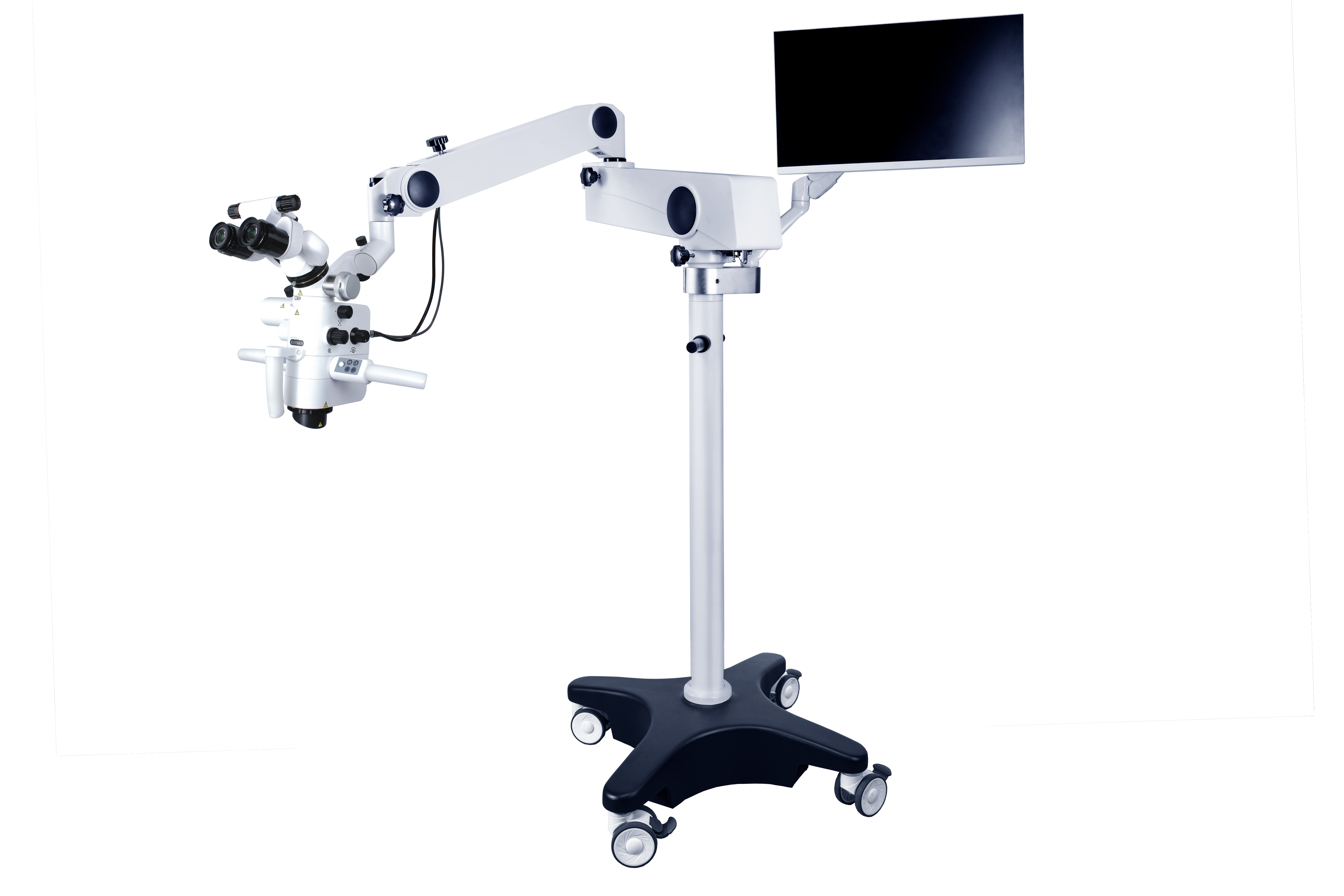
A микроскопи дандонпизишкӣаз системаи равшанӣ, системаи калонкунӣ, системаи тасвирӣ ва лавозимоти онҳо иборат аст. Системаи калонкунӣ аз окуляр, найча, линзаи объектив, танзимкунандаи калонкунӣ ва ғайра иборат аст, ки дар маҷмӯъ калонкуниро танзим мекунанд.
Гирифтани кортМикроскопи ҷарроҳии дандонпизишкии ASOM-520-DМасалан, андозаи калонкунии окуляр аз 10 × то 15 × аст, ки андозаи маъмулан 12.5X аст ва масофаи фокусии линзаи объектив дар диапазони 200 ~ 500 мм аст. Тағйирдиҳандаи андозаи калонкунӣ ду ҳолати корӣ дорад: танзими беқадами барқӣ ва танзими дастӣ бо андозаи доимии калонкунӣ.
Системаи равшании онмикроскопи ҷарроҳӣаз ҷониби манбаи рӯшноии нахи оптикӣ таъмин карда мешавад, ки барои майдони биниш равшании параллелии дурахшонро таъмин мекунад ва дар минтақаи майдони ҷарроҳӣ сояҳо ба вуҷуд намеорад. Бо истифода аз линзаҳои дурбин, ҳарду чашмро барои мушоҳида истифода бурдан мумкин аст, ки хастагиро кам мекунад; тасвири объекти сеченака ба даст оред. Як усули ҳалли мушкилоти ёрирасон ин муҷаҳҳаз кардани оинаи ёрирасон аст, ки метавонад ҳамон намуди равшанеро, ки ҷарроҳ дорад, таъмин кунад, аммо арзиши муҷаҳҳаз кардани оинаи ёрирасон нисбатан баланд аст. Усули дигар насб кардани системаи камера дар микроскоп, пайваст кардани он ба экрани намоиш ва имкон додан ба ёрирасон имкон медиҳад, ки дар экран тамошо кунанд. Тамоми раванди ҷарроҳӣ инчунин метавонад аксбардорӣ ё сабт карда шавад, то сабтҳои тиббӣ барои таълим ё тадқиқоти илмӣ ҷамъоварӣ карда шаванд.
Ҳангоми табобати бемориҳои устухон ва ҷигар,микроскопҳои ҷарроҳии дандонпизишкӣметавонад барои омӯзиши сӯрохиҳои канали реша, тоза кардани каналҳои решаи калсийшуда, таъмири сӯрохиҳои девори канали реша, омӯзиши морфологияи канали реша ва самаранокии тозакунӣ, тоза кардани асбобҳои шикаста ва тӯдаҳои шикастаи канали реша ва иҷрои...микроҷарроҳӣтартиби бемориҳои периапикӣ.
Дар муқоиса бо ҷарроҳии анъанавӣ, бартариҳои микроҷарроҳӣ инҳоянд: ҷойгиркунии дақиқи нӯги реша; Резексияи ҷарроҳии анъанавии устухон диапазони васеътар дорад, ки аксар вақт аз 10 мм зиёдтар ё баробар аст, дар ҳоле ки нобудсозии устухон бо роҳи ҷарроҳии микроҷарроҳӣ диапазони хурдтар дорад, ки аз 5 мм камтар ё баробар аст; Пас аз истифодаи микроскоп, морфологияи сатҳи решаи дандон дуруст мушоҳида карда мешавад ва кунҷи нишебии буридани реша камтар аз 10° аст, дар ҳоле ки кунҷи нишебии буридани решаи анъанавӣ калонтар аст (45°); Қобилияти мушоҳидаи истмус байни каналҳои реша дар нӯги реша; Қобилияти дақиқ омода кардан ва пур кардани нӯги реша. Илова бар ин, он метавонад нишонаҳои анатомии муқаррарии макони шикастани реша ва системаи канали решаро пайдо кунад. Раванди ҷарроҳӣ метавонад барои ҷамъоварии маълумот барои мақсадҳои клиникӣ, таълимӣ ё тадқиқоти илмӣ аксбардорӣ ё сабт карда шавад. Метавон чунин мешуморид, кимикроскопҳои ҷарроҳии дандонпизишкӣарзиши хуби татбиқ ва дурнамо дар ташхис, табобат, таълим ва таҳқиқоти клиникии бемориҳои пульпаи дандон доранд.
Вақти нашр: 19 декабри соли 2024


